Large-FOV, dual-region, two-photon fluorescence lifetime imaging microscopy (LD-2P-FLIM) for multiparameter and quantitative brain function imaging.
Large-scale imaging of multiple dynamic behaviors and quantitative neurochemical concentrations with high spatiotemporal resolution is essential for understanding complex brain functions. Two-photon microscopy (TPM) is ideally suited for in vivo brain function imaging because of its high resolution and deep tissue penetration. However, conventional TPM is limited by a restricted field-of-view (FOV), an inherent trade-off between the imaging area and temporal resolution, and an insufficient amoun
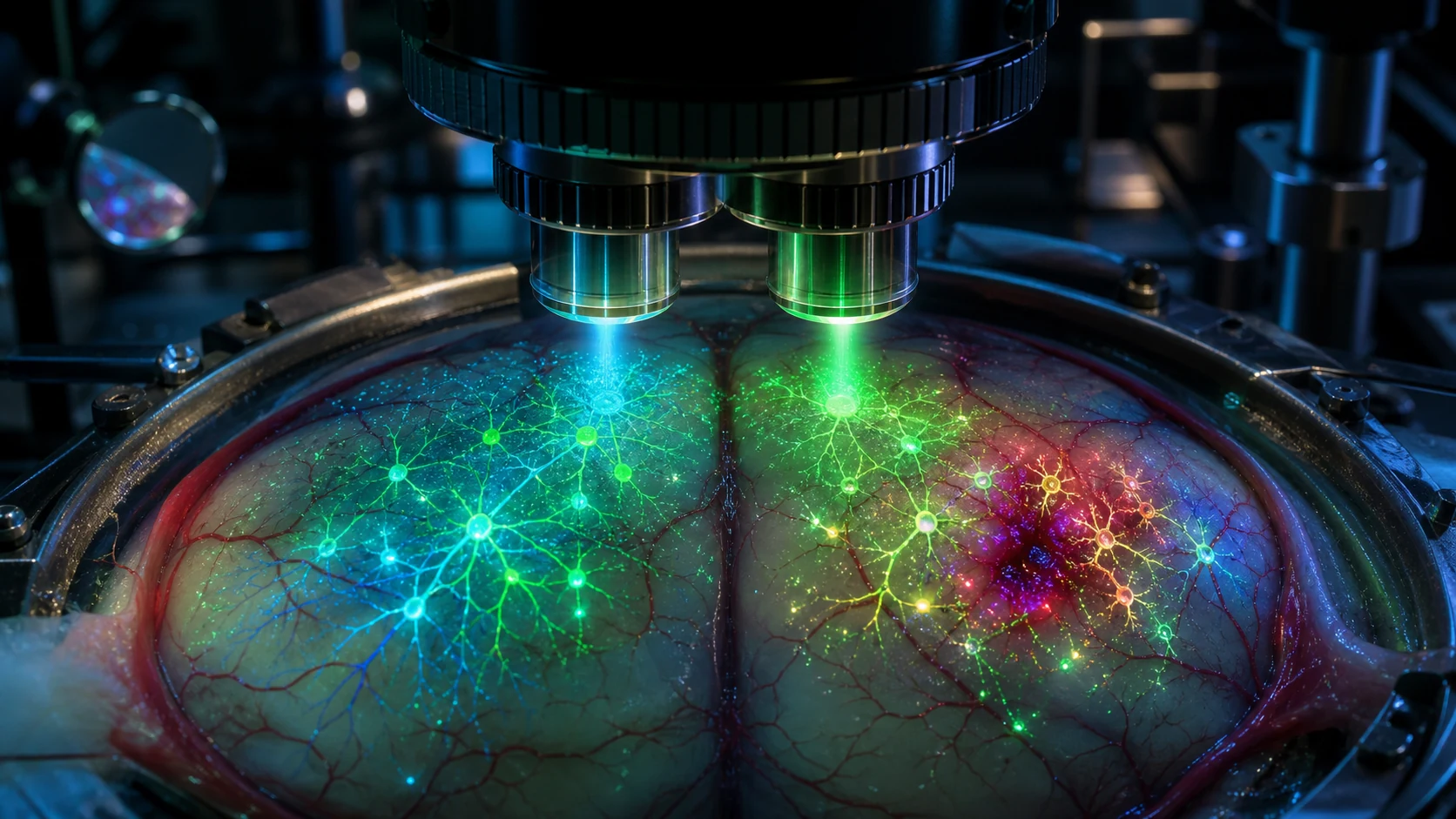
Large-scale imaging of multiple dynamic behaviors and quantitative neurochemical concentrations with high spatiotemporal resolution is essential for understanding complex brain functions. Two-photon microscopy (TPM) is ideally suited for in vivo brain function imaging because of its high resolution and deep tissue penetration. However, conventional TPM is limited by a restricted field-of-view (FOV), an inherent trade-off between the imaging area and temporal resolution, and an insufficient amount of information obtained using only intensity recording. Here, we propose large-FOV, dual-region, two-photon fluorescence lifetime imaging microscopy (LD-2P-FLIM) for multiparameter and quantitative brain function imaging, with a 3 × 3 mm 2 FOV, a uniform lateral resolution of 0.7 μm, and a FLIM throughput of up to 15.73 megapixels/s (512 × 512 pixels, 30 Hz, two regions). We extend the FOV by breaking the limit of commercial objectives with an effective adaptive optics strategy. To alleviate the trade-off between the imaging area and temporal resolution, we use a temporal multiplexing system that enables simultaneous and flexible two-region imaging across the large FOV. Furthermore, we develop a field programmable gate array module to demultiplex fluorescence signals from different regions and perform high-throughput, two-region FLIM. We demonstrate the superior performance of LD-2P-FLIM by simultaneous monitoring of neural activities across multiple cortical areas, synchronous recording of neurovascular coupling under both physiological and pathological conditions, long-term observation of the microglial response to local neuron injury, and quantitative imaging of calcium concentrations across a large neuronal population in vivo.