Ultrarapid deep 3D histology enables intraoperative mapping of glioma infiltration.
Three-dimensional (3D) histology provides volumetric insights into tissue microarchitectures across entire specimens, holding great promise for more accurate prognostication. However, existing methods are too slow for intraoperative consultations. We present ULTRA (ultrarapid cleared stimulated Raman with AI), a label-free, stain-free, fixation-free, and section-free platform that leverages the chemical specificity of stimulated Raman scattering (SRS) microscopy for rapid 3D histological analysi
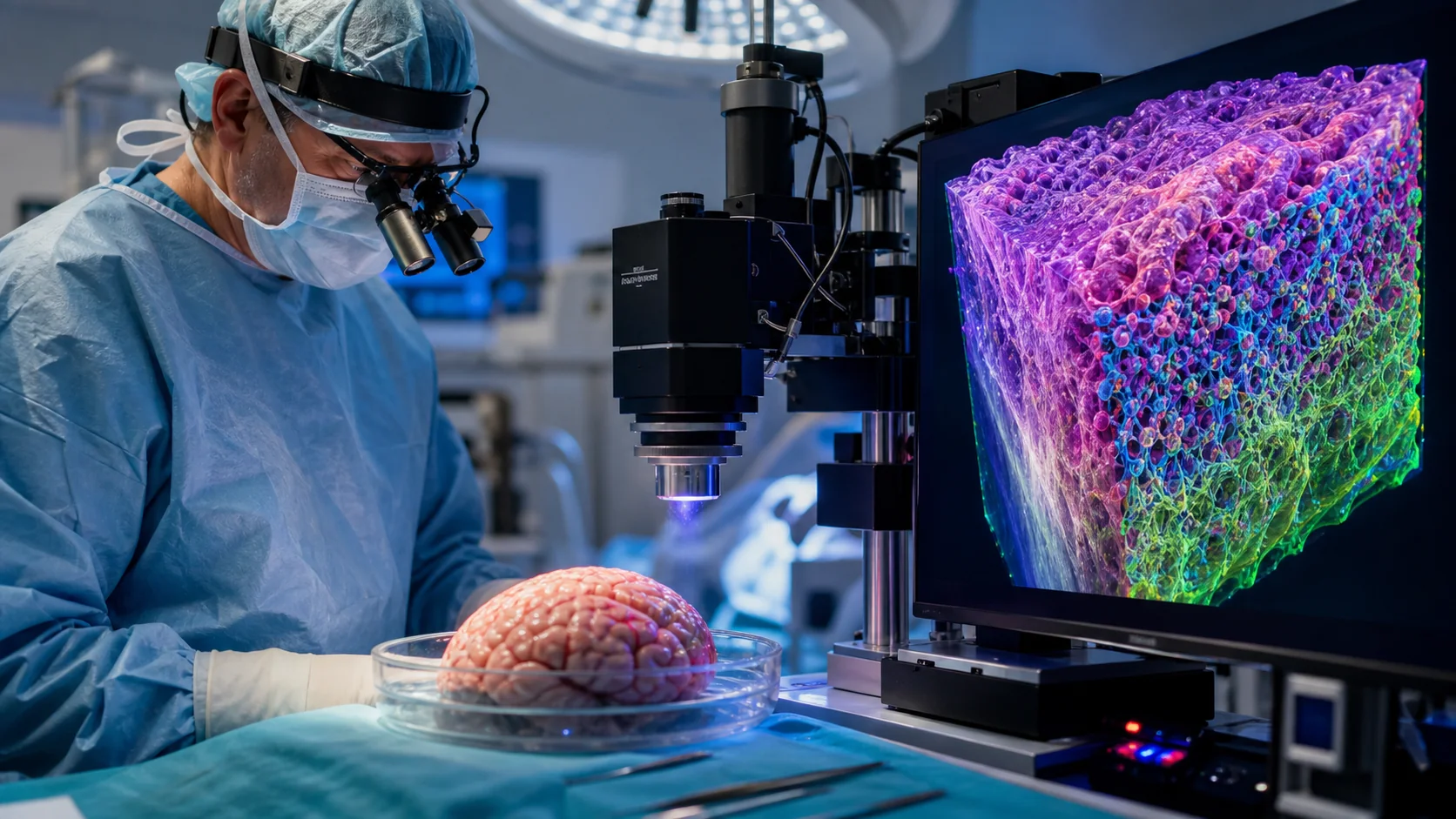
Three-dimensional (3D) histology provides volumetric insights into tissue microarchitectures across entire specimens, holding great promise for more accurate prognostication. However, existing methods are too slow for intraoperative consultations. We present ULTRA (ultrarapid cleared stimulated Raman with AI), a label-free, stain-free, fixation-free, and section-free platform that leverages the chemical specificity of stimulated Raman scattering (SRS) microscopy for rapid 3D histological analysis. Through the synergistic development of a one-step tissue-clearing protocol and unsupervised learning algorithms, ULTRA delivers high-resolution, formalin-fixed, paraffin-embedded (FFPE)-grade deep 3D virtual histology within 30 min, covering orders of magnitude more tissue than slide-based methods. In human surgical glioma samples, ULTRA accurately resolves key histological features in 3D and delineates depth-dependent tumor infiltration margins at single-cell resolution. By compressing 3D histology from days or hours to an intraoperative timescale, ULTRA addresses a critical clinical gap and enables more informed surgical decision-making in the operating room.