Normalized Raman imaging for studies of tissue physiology of the kidney.
Conventional histology relies on fixation, embedding, sectioning, and staining methods that distort cellular architecture, extract lipids, and introduce variability, limiting reproducibility. We present a pipeline for automated histopathology in the mouse kidney using normalized stimulated Raman imaging (NoRI) that enables quantitative, label-free protein and lipid measurements at high spatial resolution. NoRI computationally corrects protein, lipid, and water Raman signals, allowing quantitativ
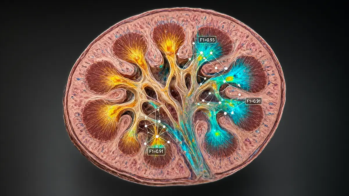
Conventional histology relies on fixation, embedding, sectioning, and staining methods that distort cellular architecture, extract lipids, and introduce variability, limiting reproducibility. We present a pipeline for automated histopathology in the mouse kidney using normalized stimulated Raman imaging (NoRI) that enables quantitative, label-free protein and lipid measurements at high spatial resolution. NoRI computationally corrects protein, lipid, and water Raman signals, allowing quantitative biomass measurements while preserving tissue architecture, facilitating analysis by convolutional neural networks (CNN) for quantitative, morphological feature extraction. Applied to mouse kidney, CNN analysis of NoRI images was able to accurately classify tubule types (F1=0.93; harmonic mean of precision and recall against immunofluorescence-defined labels), anatomical regions (F1 = 0.91), and biological sex (F1 = 0.97), revealing greater cytoplasmic lipid (+6.5 to 7.3 mg/mL; P ≤ 0.028) and nuclear protein (+26.3 mg/mL; P ≤ 0.001) in female tubules. Using acute kidney injury (AKI) in mice as a disease model to test our histopathology pipeline combining NoRI and CNNs, we captured dynamic lipid and protein organization and quantified brush border remodeling and lipid droplet changes over 25 d postinjury. Spatial lipid quantification was central for feature classification in AKI (F1 = 0.99). Quantitative NoRI images also significantly outperformed equivalent raw stimulated Raman scattering (SRS) images in distinguishing early time points (24 to 48 h) of ischemia-reperfusion injury [F1 = 0.99 (NoRI) vs. 0.84 (SRS); P < 0.0001], demonstrating the improvement of NoRI over conventional SRS imaging. These results establish our pipeline, which combines NoRI imaging with advanced CNN analyses, as a reproducible, quantitative method for histopathology and feature discovery.