Liquid Biopsies for Cancer: A Translational Science Review.
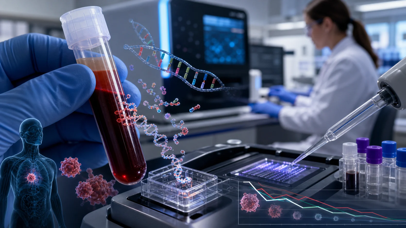
Circulating tumor DNA (ctDNA) evaluation, in which fragments of tumor DNA circulating in a patient's bloodstream are extracted and analyzed, can be used to monitor cancer progression, detect residual cancer after treatment, and identify genetic changes within cancer cells that could affect treatment response. ctDNA sequencing identifies cancer cell gene variants that inform the selection of molecularly directed therapies in several types of cancer, including non-small cell lung cancer, colorecta
Circulating tumor DNA (ctDNA) evaluation, in which fragments of tumor DNA circulating in a patient's bloodstream are extracted and analyzed, can be used to monitor cancer progression, detect residual cancer after treatment, and identify genetic changes within cancer cells that could affect treatment response. ctDNA sequencing identifies cancer cell gene variants that inform the selection of molecularly directed therapies in several types of cancer, including non-small cell lung cancer, colorectal cancer, and breast cancer. Increases or decreases in ctDNA levels can indicate treatment response (ctDNA decrease) or cancer cell resistance and recurrence (ctDNA increase). Detecting ctDNA after curative intent therapy correlates strongly with cancer recurrence and poorer survival. In a meta-analysis of 1725 patients undergoing treatment for urothelial carcinoma, higher ctDNA levels were associated with poorer survival outcomes (hazard ratio for disease-free survival, 20.69 [95% CI, 9.63-44.43]; P < .001). This association was also observed in adjuvant settings (hazard ratio for disease-free survival, 4.51 [95% CI, 3.04-6.69]; P < .001) and in patients undergoing systemic therapy for metastatic disease (hazard ratio for overall survival, 2.0 [95% CI, 1.25-3.38]; P = .004; absolute rates not available). ctDNA detection may indicate minimal residual disease, defined as cancer cells detectable only by highly sensitive testing (eg, detection of 1 cancer cell in a population of 1 million normal cells) before disease progression is identified with imaging. Detecting an early increase in ctDNA and/or a novel sequence variation that may confer resistance to standard treatment can guide therapeutic decisions, such as changing to a new treatment, before tumor progression is detectable with conventional imaging. In a prospective cohort study of 130 patients with colorectal cancer, molecular relapse of disease was detected approximately 8.7 months earlier compared with standard-of-care imaging surveillance (5.5 months vs 14.2 months; P < .001). Similarly, patients with undetectable ctDNA levels may be able to discontinue therapy and be monitored, preventing potentially unnecessary exposure to chemotherapy that may have substantial adverse effects. However, the optimal timing of ctDNA testing, management of positive results in the absence of radiographic disease, and the cost-effectiveness of serial monitoring remain unclear. ctDNA, consisting of small DNA fragments from cancer cells that can be analyzed in human blood, can help clinicians monitor cancer progression, detect minimal residual cancer, and identify genetic variants that may help guide treatment decisions. Use of ctDNA may help select best treatment and timing of therapy for a patient with cancer, but optimal clinical applications remain unclear.