Retinotopic remapping of the visual system in deaf adults.
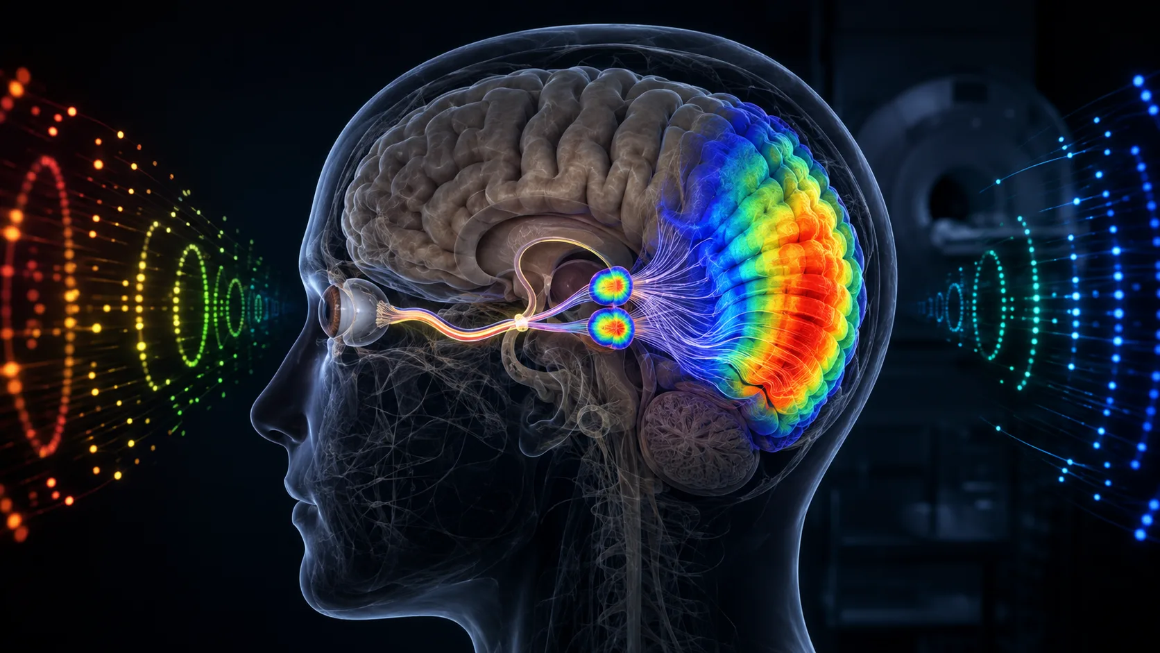
The absence of input from one sense can lead to a greater emphasis on the remaining senses, resulting in heightened performance. Numerous behavioral studies demonstrate that adults who have been deaf from an early age have superior visual sensitivity, particularly to far-peripheral stimuli. We asked whether the increased demand on peripheral vision throughout development that comes with early, profound deafness might be reflected in changes in early visual brain structures. Using functional MRI,
The absence of input from one sense can lead to a greater emphasis on the remaining senses, resulting in heightened performance. Numerous behavioral studies demonstrate that adults who have been deaf from an early age have superior visual sensitivity, particularly to far-peripheral stimuli. We asked whether the increased demand on peripheral vision throughout development that comes with early, profound deafness might be reflected in changes in early visual brain structures. Using functional MRI, we mapped visual field representations in 16 early, profoundly D/deaf adults and 16 hearing age-matched controls. D/deaf individuals exhibited a larger representation of the far-peripheral visual field in both the primary visual cortex and the lateral geniculate nucleus of the thalamus. Importantly, this was not due to a total expansion of the visual map, as there was no difference between groups in overall size of either structure, but a smaller representation of the central visual field in the D/deaf group, suggesting a redistribution of neural resources. Here, we demonstrate that the demands placed on vision due to lifelong deafness can sculpt first level visual map input, to increase neural processing resources for far-peripheral visual stimuli.