Quantitative nanoscale imaging shows peptide-MHC I complexes are monomeric and spatially regulated in human dendritic cells.
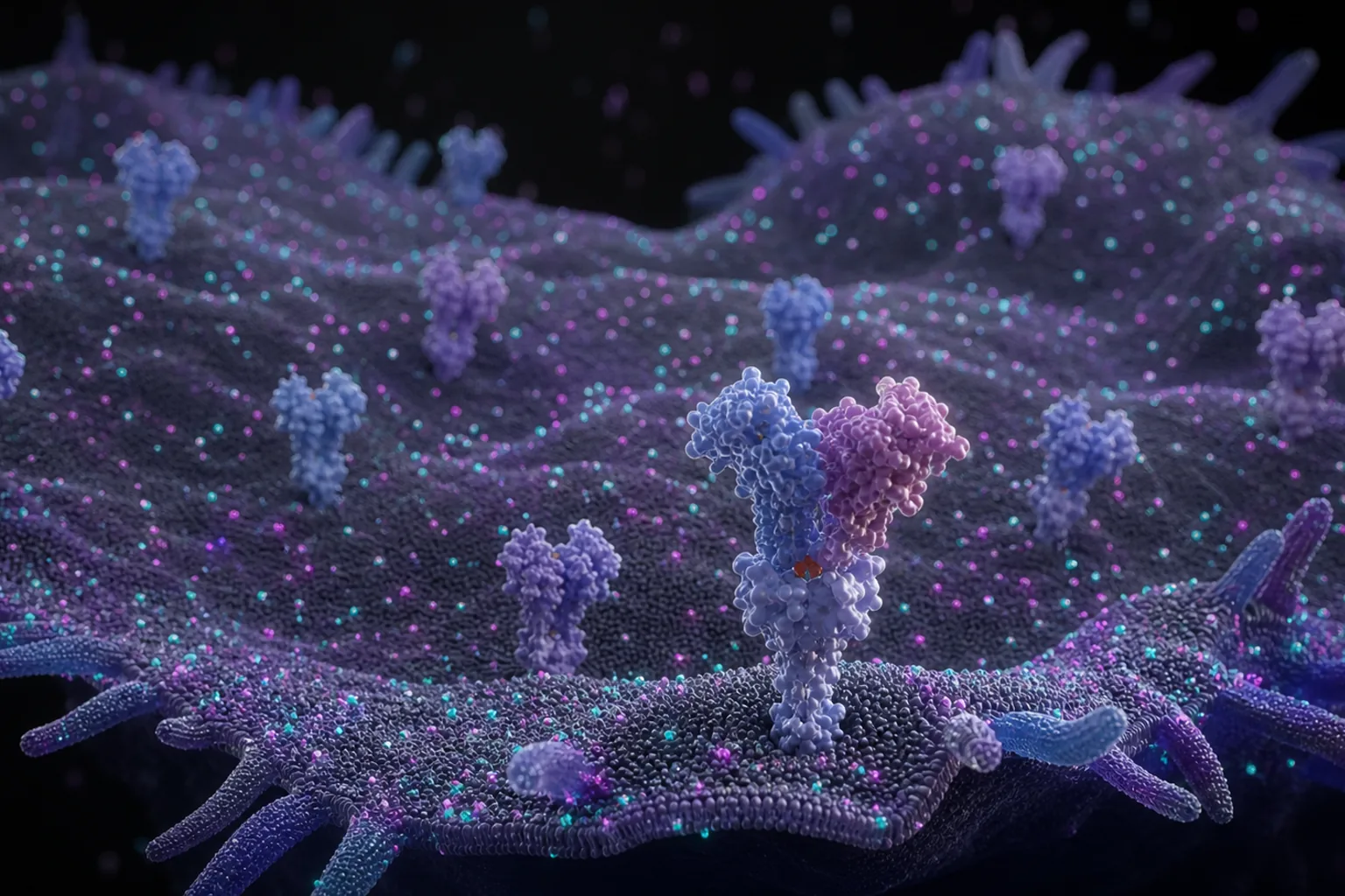
Major histocompatibility complex class I (MHC I) molecules present antigenic peptides to cytotoxic T cells, a process central to immune surveillance. However, the nanoscale spatial organization of peptide-MHC I (pMHC I) on human dendritic cells (DCs), key initiators of cytotoxic T cell responses, remains largely unexplored. Here, we combine high-affinity soluble T cell receptors with DNA-based point accumulation for imaging in nanoscale topography (DNA-PAINT) to quantitatively map and count defi
Major histocompatibility complex class I (MHC I) molecules present antigenic peptides to cytotoxic T cells, a process central to immune surveillance. However, the nanoscale spatial organization of peptide-MHC I (pMHC I) on human dendritic cells (DCs), key initiators of cytotoxic T cell responses, remains largely unexplored. Here, we combine high-affinity soluble T cell receptors with DNA-based point accumulation for imaging in nanoscale topography (DNA-PAINT) to quantitatively map and count defined pMHC I complexes at single-molecule resolution on HLA-A*02:01-expressing cells and primary human monocyte-derived DCs. We found no evidence for higher-order pMHC I nanoclusters under conditions of extracellular peptide exchange or physiological intracellular loading. Instead, detected signals correspond to individual pMHC I complexes. Notably, DC differentiation and activation modulate pMHC I surface abundance and spatial compartmentalization. These findings refine current models of antigen presentation by emphasizing regulation through surface density and spatial distribution, and establish a quantitative framework for epitope-specific, single-molecule quantification of antigen presentation in human immune cells.