Health & Medicine

Initiation of rotational collective migration in Drosophila through tissue geometry and mechanochemical feedback.
| Source: Proceedings of the National Academy of Sciences of the United States of America

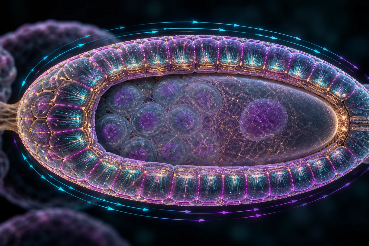
Collective migration of epithelial cells drives diverse tissue remodeling processes. In many cases, a free tissue edge works alone or in combination with other external cues to align the cells for collective movement, but how edge-free or closed epithelia become polarized for directed migration without these cues is unclear. Here, we use the rotational migration of the follicular epithelial cells in the Drosophila egg chamber to explore how cells in an edgeless epithelium initiate rotational col

Collective migration of epithelial cells drives diverse tissue remodeling processes. In many cases, a free tissue edge works alone or in combination with other external cues to align the cells for collective movement, but how edge-free or closed epithelia become polarized for directed migration without these cues is unclear. Here, we use the rotational migration of the follicular epithelial cells in the Drosophila egg chamber to explore how cells in an edgeless epithelium initiate rotational collective migration, and how the rotational axis is specified. By employing methods for long-term live imaging and delaying the onset of rotation, we show that symmetry breaking can occur at multiple developmental stages and that the atypical cadherin Fat2 promotes local motility at the basal epithelial surface before rotation begins. We then combine experiments with theoretical modeling to identify a positive feedback loop in which planar polarization of Fat2 aligns the front-rear axes of the individual cells in a common direction and the resulting tissue motion leads to the planar polarization of Fat2. This mechanosensitive feedback, coupled with rigid-body dynamics of the egg chamber, can break chiral symmetry and produce sustained rotation in silico. We further propose that mechanical constraints arising from intertissue interactions and tissue geometry ensure that rotation occurs around the anterior-posterior axis. Our findings suggest a biophysical mechanism-combining Fat2-mediated velocity-polarity alignment, rigid-body dynamics, and tissue geometry-by which a closed epithelial tissue can self-organize into persistent, large-scale rotational migration in vivo, expanding current flocking theories.

Read the original source →

Related Topics

Related Stories

Health & Medicine

PLASTID ENVELOPE ION CHANNELS (PEC1/2) link Ca 2+ and jasmonic acid signaling in plant cells.

Plants constantly encounter adverse environmental interactions. One organelle is particularly specialized in stress signaling and phytohormone synthesis: the plastid. Calcium (Ca 2+ ), a key second messenger, is known to intersect with cellular phytohormone signaling networks. While cytosolic Ca 2+ dynamics have been studied extensively, the physiological relevance of stromal Ca 2+ transients and the identity of channels mediating rapid Ca 2+ flux into plastids remain largely unexplored. In this

Continue reading
Health & Medicine

Detection of "hidden" mitotic crossovers by long-read DNA sequencing.

Mitotic crossovers in diploid organisms are usually detected by looking for loss of heterozygosity (LOH) for a marker. Assuming the crossover occurs between two duplicated chromatids, a single reciprocal crossover will result in two recombinant chromatids and two nonrecombinant chromatids. LOH will only be observed when a recombinant chromatid cosegregates with a nonrecombinant chromatid. Cells containing both recombinant chromosomes will not result in LOH. Below, we use long-range DNA sequencin

Continue reading
Health & Medicine

Amino acid homeostasis by CORVET/HOPS: A metabolic and stress resilience checkpoint for T cells.

Amino acid sufficiency is critical for T cell metabolic reprogramming, yet how T cells maintain amino acid homeostasis remains poorly defined. Here, we identify the CORVET and HOPS (CORVET/HOPS) tethering complexes as essential upstream regulators. In activated T cells, they sustain intracellular amino acid levels by promoting macropinocytosis to acquire extracellular nutrients. This function enables dual signaling outcomes: suppression of the integrated stress response (ISR) and activation of m

Continue reading
Health & Medicine

Bright monomeric fluorescent protein elite-niRFP704 for two-channel near-infrared STED nanoscopy.

The near-infrared (NIR) spectral region is attractive for live-cell imaging, due to low autofluorescence and reduced phototoxicity. Some phytochrome-derived fluorescent proteins absorb and emit fluorescence in the NIR, but have short fluorescence lifetimes and relatively low quantum yields, requiring higher laser powers thus limiting their usefulness for live-cell superresolution microscopy. Using the bacterial phytochrome miRFP703 as a template, we screened for variants with longer fluorescence

Continue reading
Health & Medicine

Quantitative nanoscale imaging shows peptide-MHC I complexes are monomeric and spatially regulated in human dendritic cells.

Major histocompatibility complex class I (MHC I) molecules present antigenic peptides to cytotoxic T cells, a process central to immune surveillance. However, the nanoscale spatial organization of peptide-MHC I (pMHC I) on human dendritic cells (DCs), key initiators of cytotoxic T cell responses, remains largely unexplored. Here, we combine high-affinity soluble T cell receptors with DNA-based point accumulation for imaging in nanoscale topography (DNA-PAINT) to quantitatively map and count defi

Continue reading