Atlas of lysosomal aging reveals a metabolite signature shared with lysosomal storage disorders
Lysosomal dysfunction is a well-recognized feature of aging. Here, we used a suite of tools for rapid lysosomal isolation to construct a multitissue atlas of the metabolite changes lysosomes undergo during aging. Aged lysosomes in brain, heart, muscle, and white adipose tissue accumulated glycerophosphodiesters and cystine, metabolites that are causally linked to juvenile lysosomal storage disorders, Batten disease, and cystinosis. Levels of these metabolites increased linearly with age, precedi
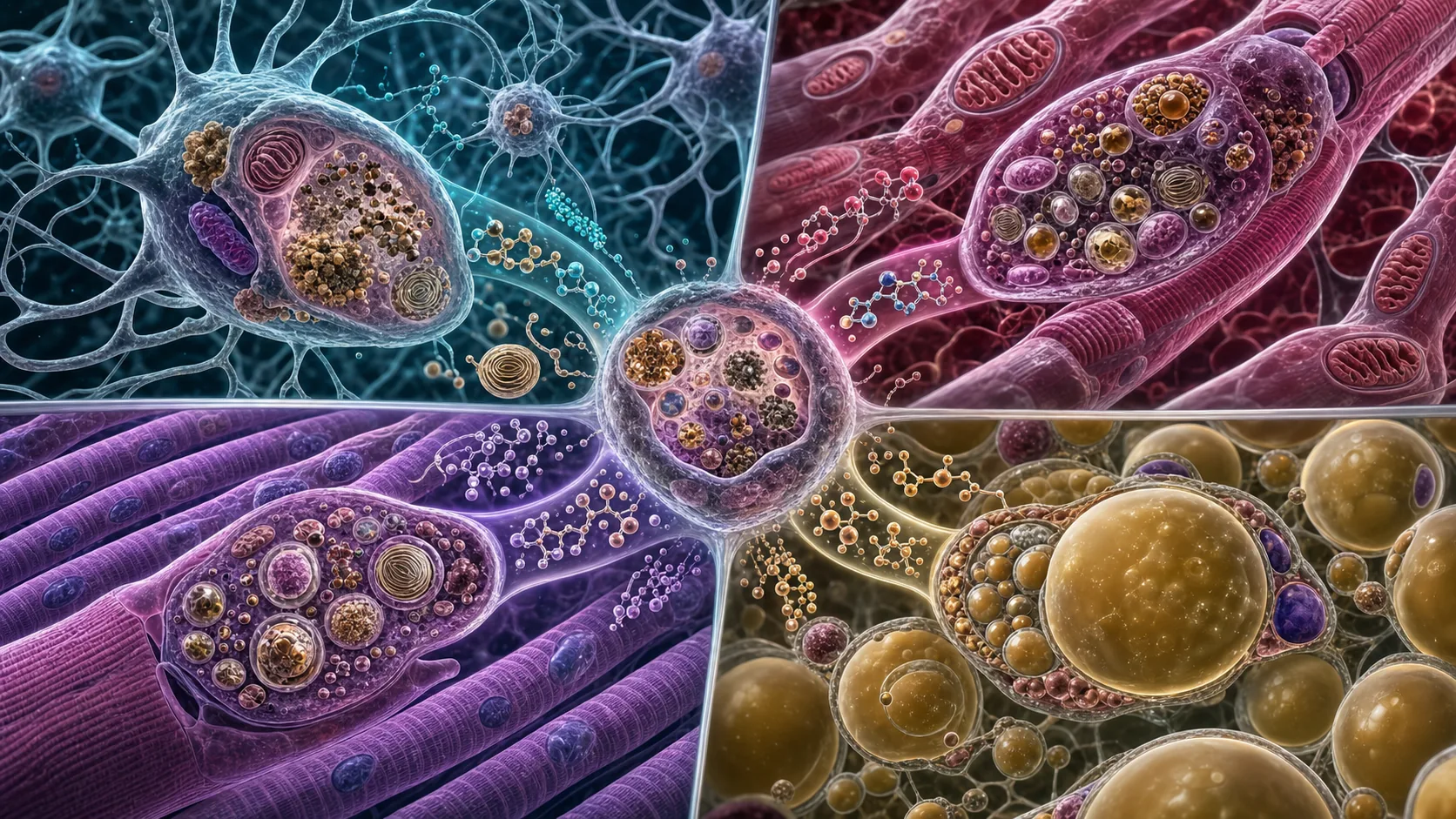
Lysosomal dysfunction is a well-recognized feature of aging. Here, we used a suite of tools for rapid lysosomal isolation to construct a multitissue atlas of the metabolite changes lysosomes undergo during aging. Aged lysosomes in brain, heart, muscle, and white adipose tissue accumulated glycerophosphodiesters and cystine, metabolites that are causally linked to juvenile lysosomal storage disorders, Batten disease, and cystinosis. Levels of these metabolites increased linearly with age, preceding organismal decline. Caloric restriction, a lifespan-extending intervention, mitigated these changes in the heart and muscle but not the brain. Our findings link lysosomal storage disorders to aging-related dysfunction and open avenues for the mechanistic investigation of how lysosomal functions deteriorate during aging and in age-associated diseases.