Health & Medicine

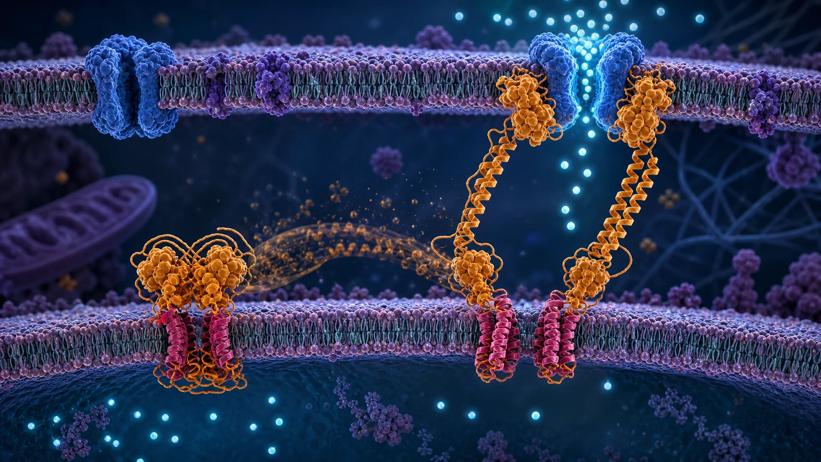
Structural rearrangements underlying the activation of STIM1 by ER calcium depletion.
| Source: Proceedings of the National Academy of Sciences of the United States of America

In resting cells, STIM1, the dimeric endoplasmic reticulum (ER) Ca 2+ sensor that controls store-operated Ca 2+ entry (SOCE), is held in a Ca 2+ -bound inactive state by multiple intramolecular restraints, or brakes. Receptor-evoked release of Ca 2+ from the ER causes a large conformational change in STIM1 that releases the brakes and exposes the CRAC activation domain (CAD), enabling it to bind and open store-operated Orai1 channels in the plasma membrane. We performed single-molecule Förs

In resting cells, STIM1, the dimeric endoplasmic reticulum (ER) Ca 2+ sensor that controls store-operated Ca 2+ entry (SOCE), is held in a Ca 2+ -bound inactive state by multiple intramolecular restraints, or brakes. Receptor-evoked release of Ca 2+ from the ER causes a large conformational change in STIM1 that releases the brakes and exposes the CRAC activation domain (CAD), enabling it to bind and open store-operated Orai1 channels in the plasma membrane. We performed single-molecule Förster resonance energy transfer (smFRET) measurements with purified STIM1 to better understand how Ca 2+ release from the luminal domain of STIM1 drives the conformational changes in the cytosolic domain that underlie CAD release. We find that Ca 2+ removal releases the CAD from CC1α1 (the "CC1 clamp") without obligatory formation of the CC1 coiled-coil that has been associated with CAD release in cells. Surprisingly, the CAD rearranges dramatically during release, as the two hairpin protomers that create its characteristic V-shaped structure are spread apart. Locking the two protomers together by cysteine crosslinking prevents CAD release, suggesting that the CAD must rearrange to escape the CC1 clamp. Our data support a model in which ER depletion-induced dimerization of the luminal SAM domains drives the cytosolic domain into multiple intermediate states including a 3-helix bundle of CC1α1/2/3, releasing the CC1 clamp and allowing the CAD to escape through a "fold-out" mechanism. Subsequent formation of the CC1 coiled-coil enables the CAD to revert to its original shape and extends it toward the plasma membrane to activate Orai1.

Read the original source →

Related Stories

Health & Medicine

PLASTID ENVELOPE ION CHANNELS (PEC1/2) link Ca 2+ and jasmonic acid signaling in plant cells.

Plants constantly encounter adverse environmental interactions. One organelle is particularly specialized in stress signaling and phytohormone synthesis: the plastid. Calcium (Ca 2+ ), a key second messenger, is known to intersect with cellular phytohormone signaling networks. While cytosolic Ca 2+ dynamics have been studied extensively, the physiological relevance of stromal Ca 2+ transients and the identity of channels mediating rapid Ca 2+ flux into plastids remain largely unexplored. In this

Continue reading
Health & Medicine

Detection of "hidden" mitotic crossovers by long-read DNA sequencing.

Mitotic crossovers in diploid organisms are usually detected by looking for loss of heterozygosity (LOH) for a marker. Assuming the crossover occurs between two duplicated chromatids, a single reciprocal crossover will result in two recombinant chromatids and two nonrecombinant chromatids. LOH will only be observed when a recombinant chromatid cosegregates with a nonrecombinant chromatid. Cells containing both recombinant chromosomes will not result in LOH. Below, we use long-range DNA sequencin

Continue reading
Health & Medicine

Amino acid homeostasis by CORVET/HOPS: A metabolic and stress resilience checkpoint for T cells.

Amino acid sufficiency is critical for T cell metabolic reprogramming, yet how T cells maintain amino acid homeostasis remains poorly defined. Here, we identify the CORVET and HOPS (CORVET/HOPS) tethering complexes as essential upstream regulators. In activated T cells, they sustain intracellular amino acid levels by promoting macropinocytosis to acquire extracellular nutrients. This function enables dual signaling outcomes: suppression of the integrated stress response (ISR) and activation of m

Continue reading
Health & Medicine

Bright monomeric fluorescent protein elite-niRFP704 for two-channel near-infrared STED nanoscopy.

The near-infrared (NIR) spectral region is attractive for live-cell imaging, due to low autofluorescence and reduced phototoxicity. Some phytochrome-derived fluorescent proteins absorb and emit fluorescence in the NIR, but have short fluorescence lifetimes and relatively low quantum yields, requiring higher laser powers thus limiting their usefulness for live-cell superresolution microscopy. Using the bacterial phytochrome miRFP703 as a template, we screened for variants with longer fluorescence

Continue reading
Health & Medicine

Quantitative nanoscale imaging shows peptide-MHC I complexes are monomeric and spatially regulated in human dendritic cells.

Major histocompatibility complex class I (MHC I) molecules present antigenic peptides to cytotoxic T cells, a process central to immune surveillance. However, the nanoscale spatial organization of peptide-MHC I (pMHC I) on human dendritic cells (DCs), key initiators of cytotoxic T cell responses, remains largely unexplored. Here, we combine high-affinity soluble T cell receptors with DNA-based point accumulation for imaging in nanoscale topography (DNA-PAINT) to quantitatively map and count defi

Continue reading