Health & Medicine

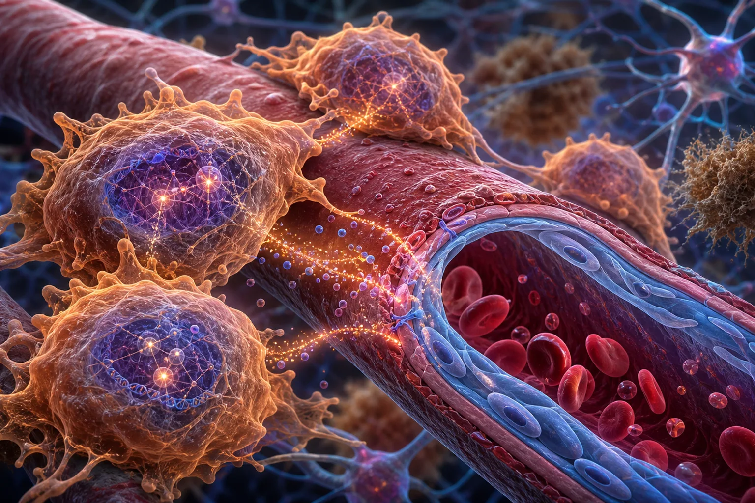
Brain perivascular macrophages regulate endothelial cell function via a cMAF-dependent transcriptional program in mouse and human.
| Source: Cell

Brain perivascular macrophages maintain brain physiology, yet their transcriptional regulators and functions in health and disease remain unclear. Using single-cell multi-omics and functional experiments, we identify cellular musculoaponeurotic fibrosarcoma oncogene (cMAF) as a key transcription factor for brain perivascular macrophages, and conditional deletion of cMAF disrupts their phenotype in vivo. Functionally, cMAF drives insulin-like growth factor-1 (IGF1) expression in perivascular macr

Brain perivascular macrophages maintain brain physiology, yet their transcriptional regulators and functions in health and disease remain unclear. Using single-cell multi-omics and functional experiments, we identify cellular musculoaponeurotic fibrosarcoma oncogene (cMAF) as a key transcription factor for brain perivascular macrophages, and conditional deletion of cMAF disrupts their phenotype in vivo. Functionally, cMAF drives insulin-like growth factor-1 (IGF1) expression in perivascular macrophages, enabling communication with endothelial cells. Consistently, cMAF deletion in perivascular macrophages causes transcriptional alterations in cerebral arteries, affecting vascular functions. Notably, cMAF emerges as the main transcription factor for human perivascular macrophages, suggesting conservation of this transcriptional module. During Alzheimer's disease (AD), human perivascular macrophages upregulate cMAF and IGF1 to enhance communication with vascular cells, and this response is abrogated in APOE4 carriers. Lastly, we explore an uncharacterized polymorphism in cMAF, providing evidence that the cMAF program is protective against AD. Targeting cMAF in perivascular macrophages may offer new therapeutic strategies for neurodegenerative and cerebrovascular diseases.


Read the original source →

Related Stories

Health & Medicine

PLASTID ENVELOPE ION CHANNELS (PEC1/2) link Ca 2+ and jasmonic acid signaling in plant cells.

Plants constantly encounter adverse environmental interactions. One organelle is particularly specialized in stress signaling and phytohormone synthesis: the plastid. Calcium (Ca 2+ ), a key second messenger, is known to intersect with cellular phytohormone signaling networks. While cytosolic Ca 2+ dynamics have been studied extensively, the physiological relevance of stromal Ca 2+ transients and the identity of channels mediating rapid Ca 2+ flux into plastids remain largely unexplored. In this

Continue reading
Health & Medicine

Detection of "hidden" mitotic crossovers by long-read DNA sequencing.

Mitotic crossovers in diploid organisms are usually detected by looking for loss of heterozygosity (LOH) for a marker. Assuming the crossover occurs between two duplicated chromatids, a single reciprocal crossover will result in two recombinant chromatids and two nonrecombinant chromatids. LOH will only be observed when a recombinant chromatid cosegregates with a nonrecombinant chromatid. Cells containing both recombinant chromosomes will not result in LOH. Below, we use long-range DNA sequencin

Continue reading
Health & Medicine

Amino acid homeostasis by CORVET/HOPS: A metabolic and stress resilience checkpoint for T cells.

Amino acid sufficiency is critical for T cell metabolic reprogramming, yet how T cells maintain amino acid homeostasis remains poorly defined. Here, we identify the CORVET and HOPS (CORVET/HOPS) tethering complexes as essential upstream regulators. In activated T cells, they sustain intracellular amino acid levels by promoting macropinocytosis to acquire extracellular nutrients. This function enables dual signaling outcomes: suppression of the integrated stress response (ISR) and activation of m

Continue reading
Health & Medicine

Bright monomeric fluorescent protein elite-niRFP704 for two-channel near-infrared STED nanoscopy.

The near-infrared (NIR) spectral region is attractive for live-cell imaging, due to low autofluorescence and reduced phototoxicity. Some phytochrome-derived fluorescent proteins absorb and emit fluorescence in the NIR, but have short fluorescence lifetimes and relatively low quantum yields, requiring higher laser powers thus limiting their usefulness for live-cell superresolution microscopy. Using the bacterial phytochrome miRFP703 as a template, we screened for variants with longer fluorescence

Continue reading
Health & Medicine

Quantitative nanoscale imaging shows peptide-MHC I complexes are monomeric and spatially regulated in human dendritic cells.

Major histocompatibility complex class I (MHC I) molecules present antigenic peptides to cytotoxic T cells, a process central to immune surveillance. However, the nanoscale spatial organization of peptide-MHC I (pMHC I) on human dendritic cells (DCs), key initiators of cytotoxic T cell responses, remains largely unexplored. Here, we combine high-affinity soluble T cell receptors with DNA-based point accumulation for imaging in nanoscale topography (DNA-PAINT) to quantitatively map and count defi

Continue reading